Histotripsy: Making the Impossible Possible
By Anna Megdell Photos by Erica Reist Bass
A surprising discovery in graduate school led U-M biomedical engineer Zhen Xu to the first non-invasive, non-thermal tumor resection technique using ultrasound. Twenty years later, she and a team of Rogel researchers have received FDA approval to treat patients with liver cancer. Here’s how they did it.
In 2002, Zhen Xu, Ph.D., was a graduate student at the University of Michigan searching for a research project. Her adviser, Charles Cain, Ph.D., founding chair and professor of biomedical engineering,, was approached by a pediatric cardiologist, Achi Ludomirsky M.D., who asked Cain and Xu if they could develop a approach to treat children with congenital heart disease by resecting heart tissue without cutting open the patient to put further strain on already sick kids. The procedure needed to be non-invasive and non-toxic. After months of research, nothing had worked.
“Cain told me that I couldn’t rely on preexisting research or literature. I was trying to make something completely new. He said I needed to think ‘out-of-the-box,’” says Xu, now a professor of biomedical engineering at the U-M College of Engineering. “His exact words were, ‘I don't want your innovative mind to be contaminated by conventional wisdom.’ I thought ‘that’s great, but how!?’”
Xu began investigating how ultrasounds generate cavitation, a process that creates micro-bubbles in tissue that expand and collapse, eventually erasing the tissue. Ultrasound was known to be non-toxic and non-invasive; babies underwent imaging all the time at no harm to them, so Xu figured it could be effective. “But at the time, nobody thought cavitation could be controlled to actually target and remove specific tissue,” she explains.
For months, Xu explored new ways to manipulate ultrasounds to no avail. Even though ultrasound is above that human hearing range, ultrasound bursts to generated cavitation is typically pulsed at a frequency that human can hear, including her lab mates, could hear. “They said my experiment was too loud,” Xu says, laughing. Then one day, using pig hearts, Xu tested how tissue responded when submerged in water and exposed to pulsing sound waves at a very powerful amplitude, but with very short bursts of microsecond length and pulsed at a frequency beyond the human hearing range.
“It only took a few seconds,” she says. “I saw something that looked like smoke rising out of the water. But it turns out it wasn’t smoke. It was tiny, tiny debris from the tissue.
“In one minute, I saw a hole in the pig heart tissue,” Xu continues. “My first reaction was, ‘Am I dreaming?’”
Science Behind the Serendipity
Photos by Erica Reist Bass
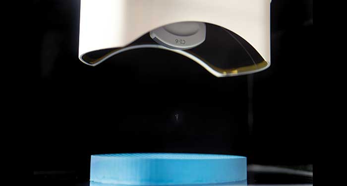
This breakthrough led Xu down a decades-long path to developing what is now known as histotripsy, a completely non-invasive—no needles, no incision, no exposure to radiation, non-thermal—way to destroy the target tissue. Instead of traditional methods, histotripsy functions mechanically. Ultrasound waves are pinpointed at inside the target tissue or a tumor to generate a cluster of microbubbles from the pre-existing nano-meter gas pockets. Microbubbles contract and expand repeatedly adjacent to cells, producing high local mechanical strain on the cells. Eventually, the cell walls get destroyed. Because it’s mechanical, the cell death is irreversible.
But before histotripsy was fully understood, Xu needed to make sure the hole she saw in the tissue wasn’t a fluke. She repeated the process three times, in different locations, and each time, a hole emerged. “Then I could finally let myself admit that what I saw was true,” Xu says. She showed her results to Cain. “We realized we had something special on our hands. This was the beginning of our journey.”
From there, Xu and a team began to study which parameters led to the hole in the tissue. She knew she could target and destroy tissue using ultrasound; now she needed to understand how it happened. “I was able to recreate it in terms of engineering,” Xu explains. “But it wasn’t clear why those specific parameters had worked. We needed to understand the precise mechanism on how cavitation was generated and controlled so we could tell the scientific community that why, in the past five decades, nobody was able to generate and control cavitation to treat patients, but now we could.”
Xu admits that the first discovery, the very first hole in the tissue, was serendipity. “I tried some parameters that were totally different from anything I tried before, and it worked out.” From there came the hard work, and many years, of scientific exploration to turn the initial discovery into a real technology that could eventually be used on patients.
For a decade, through graduate school and into her post-doctoral and early career years, Xu and team members studied the mechanisms behind histotripsy and developed the equipment needed to make results consistent. “Our job is to make the impossible possible,” she says. “I find inspiration when it looks like things can’t be accomplished. It makes me want to look closer.”
Once Xu and her team established the mechanics underlying histotripsy, she reached out to Mishal Mendiratta-Lala, M.D., and Clifford Cho, M.D., at Michigan Medicine to begin exploring if this new technology could be used clinically to help patients with tumors.
Building the Team

Photos by Erica Reist Bass
In 2017, Mendiratta-Lala, professor of radiology, heard about a researcher in biomedical engineering who was looking for someone to help interpret liver images on animals. “Right away, I was very interested in getting involved,” she says. That researcher was Xu. Together, they started a preclinical project to see if histotripsy worked on liver tumors in animal models.
Xu also reached out to Cho, professor of surgery, who runs a lab that studies cancer immunotherapy.
“We did a bunch of experiments that showed histotripsy could be used as a platform for cancer immunotherapy.”
These projects led to more research, which led to collaborating with researchers from the University of Wisconsin. This preclinical worked helped establish a clinical trial to test histotripsy on human patients who have either primary or metastatic liver cancer in the United States and Europe.
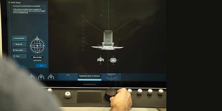
The trial’s run-of-show is straightforward. Patients get an MRI within 30 days of enrollment and standard blood and liver tests the week of the procedure. On the day of the procedure, they go under general anesthesia to control their breathing. Radiologists perform the histotripsy procedure in the interventional radiology procedure room. They get an immediate post-procedure imaging study to confirm that the tumor was completely treated. After the procedure, the patient stays in recovery until the anesthesia wears off. They then can go home that same day, returning for imaging a month later to make sure there is no disease recurrence.
The results of the clinical trial showed histotripsy was safe and effective in destroying liver tumors and led to complete tumor regression.
But the clinical trial results also revealed another effect: Not only did histotripsy destroy the targeted area of the organ, but it triggered the body’s immune system as well. Cho explains that common cancer treatments don’t often effectively incite an anti-tumor immune response. Many tumor ablation techniques rely on methods like heating or freezing the tumor, which prevents the immune system’s ability to see and recognize the tumor. But histotripsy’s mechanical nature changes the game.
“A lot of our preliminary studies demonstrated, in various forms in many kinds of cancers, that whenever we ablated the tumors using histotripsy in mice, we saw very measurable immune responses generated against that tumor,” Cho explains. “For example, if there were two tumors in mice and we used histotripsy on just one, we saw an anti-tumor immune response against the other tumor.”
“We noticed that if we treat with histotripsy, the whole tumor as well as tumors elsewhere in that animal were going away a few weeks later,” Mendiratta-Lala adds. “We’re seeing this immune system activation and we're priming the body to fight tumor elsewhere. Triggering the immune system could lead to things like addressing distant metastasis or making immunotherapies more potent. If this really is going to happen in patients, it will obviously be life changing.”
Now, the team is interested in understanding why. They are conducting another clinical trial combining histotripsy and immunotherapy. “We still need to see how the immune response works in human patients,” says Mendiratta-Lala. Early data from the clinical trial in Spain showed that untreated tumors shrink concurrently with tumors that have received treatment.
In addition to destroying the tumor, results show that histotripsy changes the tumor microenvironment, altering its blood oxygen content. “It convinces tumor cells not just to die but to die in specific pathways that are almost perfect for generating immune response,” Cho explains. “We found that it actually reprogrammed immune cells to acquire certain abilities to kill tumor cells that they didn’t have before.”
For Cho, this leg of the research—investigating histotripsy’s unexpected effects on the immune system—embodies what it means to follow the science.
“These immune effects have led us to more interesting and nuanced areas of study than we ever could have anticipated.” he says.
Patients First

In addition to the clinical trial’s staggering results, the clinicians on the team emphasize what it means for patients to have a treatment that is successful and completely non-invasive.
“As someone who does interventional procedures, recovery from histotripsy versus other forms of treatment is incredible,” says Mendiratta-Lala. “Usually after an ablation procedure that uses microwave or radiofrequency, patients can have tenderness and a little bit of pain; they can’t move for five hours after the procedure.” But with histotripsy, the only recovery is from the general anesthesia. “Many patients ask if the procedure was even done because they don't feel any pain,” Mendiratta-Lala continues. “They can just get up and walk out the door when they’re done.”
Valerie Khaykin is the clinical trial coordinator of the histotripsy trial. She recruited patients to the trial and guided them through the process. U-M was the highest enrolling institution with 10 patients to date thanks to Khaykin’s efforts. She says the most surprising part of the trial for the patients lies in the consults. “Patients and sometimes even nurses and the medical professionals can’t believe there’s no recovery aside from the anesthesia,” she says. “We’ve gotten really good at explaining how the technology works.”
Khaykin says that histotripsy is especially comforting for patients who might not qualify for typical treatments. “For people in their 80s or for those who have co-morbidities, not having to undergo traditional surgery to remove their tumors is a gamechanger.”
Strict guidelines are put in place on who can qualify for the clinical trial. While necessary to prove the effectiveness of histotripsy and to establish a level baseline, the qualifications can exclude patients whose labs are just outside the established range of candidacy but who might still benefit from treatment. For Khaykin, these are the patients who stay with her, and who come to mind when thinking about how FDA approval will impact many patients with cancer.
“With FDA approval, we now won’t have to worry about those technicalities,” she says. “For some patients, histotripsy is curative. But even if it’s not, it has the potential to activate their immune system or ease disease burden. It’s a relatively benign treatment and doesn’t discriminate in terms of age or disease progression. We’ve treated so many people with different pathologies, different overall health. It has the potential to be a great, universal option. It’s giving patients a lot of hope.”
To FDA and Beyond
HistoSonics, a startup company that licensed Xu and her team’s histotripsy patents from U-M and created the clinical histotripsy device (EdisonTM), received FDA approval to use the device to treat patients with liver disease using histotripsy in October 2023. Though the culmination of 20 years of work, Xu is eager for the next step. “We’re aiming for FDA approval on renal tumors and pancreatic tumors. And there’s the combination trial between histotripsy and immunotherapy. We now have this technology that hopefully will be used for many, many things,” she says.
Xu receives emails from patients around the world, wanting to know if histotripsy could be used to help treat their disease. “I’ve been working on this for over 20 years, and there are so many people involved who have been working so hard,” she continues.
“Now we’re at the point where we can really see that it’s going to benefit patients. There are so many people who need help. It’s not time to stop. We have more work to do.”
&nbps;
Disclosure: U-M retains a financial interest in HistoSonics, as do a number of researchers who were involved in this project and who helped develop the technology licensed to HistoSonics, including Xu, who is a company co-founder, stockholder and consultant, and Cho, who is a consultant. Each stands to benefit financially from the success of the platform.